Abstract
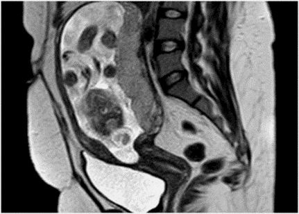
Meningo encephalocele is a congenital anomaly and is a neural tube defect with occipital meningo encephalocele being the most common and is a result of a failure of the surface ectoderm to separate from the neuroectoderm. This condition can be identified in 1st trimester in 80% of cases and almost all by 2nd trimester. A 20-year-old third gravida was referred for antenatal Ultrasonography at five months of amenorrhoea to rule out fetal anomalies. On targeted imaging, for fetal anomalies, a defect was seen in occipital bone with herniation of posterior fossa contents with overlying meningeal covering. No other fetal anomalies were noted. A diagnosis of isolated occipital meningoencephalocele was made with additional fetal MRI correlation. The mother underwent termination of her pregnancy by Department of Obstetrics and Gynecology because of the grim fetal prognosis. The mother was advised to plan the subsequent pregnancies and was advised pre-conceptional folic acid supplementation. We present a case of isolated occipital meningoencephalocele- a rare congenital anomaly which was diagnosed prenatally in our hospital. This case provides an opportunity for identifying such neurological defects early and prompt termination of pregnancy to prevent comorbidity to mother. This study also helps to establish occipital meninigioencephalocele as an isolated clinicoradiological diagnosis and to distinguish it from syndrome associated occipital meninigioencephalocele or those associated with other neural tube defects like Chiari III malformations. It also allows us to stress once again the role of periconceptional folic acid in preventing the occurrence of neural tube defects.
Full text article
Authors

This work is licensed under a Creative Commons Attribution-NonCommercial-NoDerivatives 4.0 International License.

